

Why Are X-Rays Safe for Imaging but Risky in Excess?
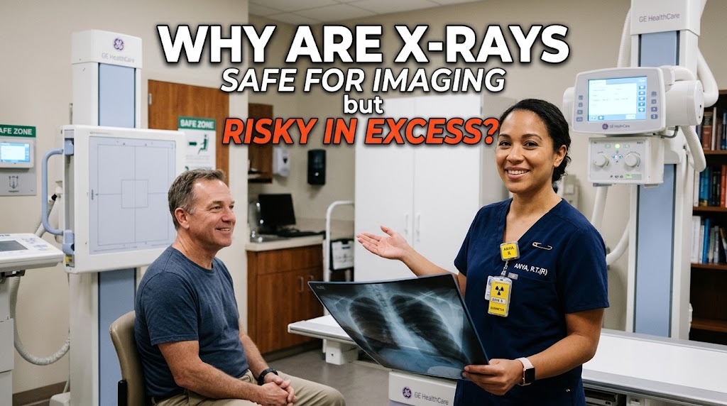
X-rays are a form of high-energy electromagnetic radiation that can easily pass through soft biological tissues. Because bones and denser materials absorb more energy, X-rays create clear contrast images of internal body structures.
In medical environments, diagnostic X-rays are delivered in very low, controlled doses. This controlled application ensures that doctors can quickly identify medical issues without causing harm to surrounding tissues.
WHY SINGLE DIAGNOSTIC DOSES ARE SAFE
A single medical X-ray exposes the body to an extremely small amount of radiation. To put it in perspective, one chest X-ray delivers roughly the same radiation dose that a person naturally absorbs from background environmental sources over a few days.
Human cells are equipped with natural repair mechanisms that efficiently fix tiny amounts of cellular damage caused by low exposures. Because the body heals rapidly from minor exposures, small diagnostic doses pose negligible risks to healthy individuals.
THE RISKS OF EXCESSIVE EXPOSURE
While low doses are safe, repeated or excessive exposure to ionizing radiation carries increased health risks. X-rays possess enough packet energy to knock electrons out of atoms, which can break chemical bonds within biological cells.
When cells sustain high amounts of energy over short periods or frequent bursts, it can lead to cellular mutations or DNA damage. Over time, unmapped mutations increase the long-term likelihood of developing abnormal cell growths or tissue damage.
HOW MEDICAL PROFESSIONALS MANAGE DOSAGE
Radiologists follow a fundamental safety principle known as ALARA (As Low As Reasonably Achievable) to minimize radiation exposure. Equipment is carefully calibrated to use the minimum required radiation intensity to produce clear, diagnostic-quality images.
Additionally, healthcare providers protect non-target body parts by using protective lead aprons and shields. By limiting the beam to only the required area, medical teams significantly reduce unnecessary exposure to nearby healthy tissues.
BENEFIT VERSUS RISK EVALUATION
Every medical procedure involving radiation involves a careful evaluation of potential benefits against possible risks. The immediate diagnostic value of detecting broken bones, infections, or internal conditions far outweighs the minimal long-term radiation risk.
Patients can maintain peace of mind by keeping an accurate personal medical imaging history. Discussing previous scans with doctors ensures that duplicate procedures are avoided, maintaining a safe balance of health monitoring.
SAFE PRACTICES IN MODERN RADIOLOGY
Modern technology continuously improves radiological safety through advanced digital imaging sensors. Digital X-ray systems require far less radiation exposure than older film-based equipment while providing higher image accuracy.
Regulatory health agencies establish strict occupational exposure limits for technicians and doctors who operate X-ray machinery daily. These combined efforts guarantee that diagnostic radiology remains a highly secure and invaluable tool in modern healthcare.
CONCLUSION
In summary, X-rays are safe for medical imaging because doses are strictly controlled, allowing natural cellular repair mechanisms to manage tiny exposures safely. However, excessive exposure carries risks because cumulative ionizing energy can disrupt cellular structures over time.
By employing protective shielding, digital imaging, and strict dosage guidelines, healthcare systems maximize diagnostic safety. Understanding this balance ensures that patients receive effective diagnostic care without unnecessary risk.
Recent Posts

Bagaimana Sistem SKS pada Program Kuliah... 22.11.2026/53 View

How Does Technology Make Daily Evaluation... 21.11.2026/82 View

Apa Strategi Menghadapi Kesenjangan antara Kompetensi... 21.11.2026/81 View

Mengapa Inventaris Aset Penting bagi Kelancaran... 21.11.2026/77 View

When Should Musicians Join a Record... 21.11.2026/82 View
